


Transient Osteoporosis of the Hip
Hip Anatomy
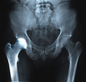
The hip joint is a ball-and-socket joint. A part of the pelvis bone known as the acetabulum forms the socket and the upper end of the femur, known as the femoral head, forms the ball.
What is Transient Osteoporosis of the Hip?
Transient osteoporosis of the hip is a rare condition that causes temporary bone loss in the upper region of the thighbone (femur). It occurs most often in young or middle-aged men of the age groups 30 to 60, and women in their later stages of pregnancy or early postpartum (following childbirth). It is characterized by abrupt onset of pain that increases with activity.
Causes of Transient Osteoporosis of the Hip
In transient osteoporosis of the hip, the femoral head loses its density and strength and becomes more prone to breaking. The exact cause for this is unknown. Some of the proposed causes include atypical mechanical stresses acting on the hip joint, hormonal abnormalities and blockage of some of the small blood vessels surrounding the hip joint.
Symptoms of Transient Osteoporosis of the Hip
The symptoms of transient osteoporosis of the hip may include:
- Unknown pain in the hip not triggered by any previous accident or injury
- Abrupt onset of pain in the anterior thigh, the side of the hip, groin or buttocks
- Pain that increases with activity or weight-bearing, and decreases with rest
- Intense pain with extreme hip range of motion
- Gradually increasing pain that becomes disabling over a few weeks or months
- A prominent limp
Diagnosis of Transient Osteoporosis of the Hip
The diagnosis of transient osteoporosis of the hip often begins with a review of your history and a physical examination. You will be asked to perform various range-of-motion exercises to replicate your pain. You may experience acute pain with weight-bearing and active range of motion and minimal pain when your doctor moves the hip for them (passive range of motion). This is one of the indicators in the diagnosis of transient osteoporosis of the hip.
You will also be recommended to undergo imaging studies such as X-rays, CT scans, MRIs, or nuclear scans to further document transient osteoporosis of your hip.
Most patients with transient osteoporosis of the hip are found to have bone marrow edema. Bone marrow edema is a condition where fluid builds up in the bone marrow (spongy material located in the hollow of the long bones) and the bone marrow becomes inflamed. MRI scans have been found to be particularly beneficial in documenting bone marrow edema and are one of the most practicable studies in the diagnosis of transient osteoporosis of the hip.
Treatments for Transient Osteoporosis of the Hip
Transient osteoporosis of the hip resolves on its own and treatment involves preventing any damage to the weakened bones and minimizing the symptoms and discomfort. Treatments include:
Medication: Non-steroidal anti-inflammatory medications or NSAIDs may be recommended to alleviate inflammation and pain
Restricted weight-bearing: You may be recommended to restrict or to completely avoid putting weight on your hip joint. You may need to use walking aids such as crutches, cane or a walker to limit the stress on your hip bone.
Physical therapy: Your doctor may instruct you on special exercises to help strengthen the muscles supporting your hip. Water exercises have been found to be helpful as they ease movement and relieve weight-bearing.
Nutrition: Vitamin D and calcium have been found to be effective in healing and rebuilding of bones. Your doctor will recommend foods or supplements that can help you recover faster.
Related Topics:
- Hip Adductor Injuries
- Pediatric Femur Fracture
- Stress Fractures of the Hip
- Avulsion Fractures of the Pelvis
- Hip Injury
- Stem Cell Therapy for Hip Injuries
- Gluteus Tendon Tear
- Hip Pain
- Snapping Hip Syndrome
- Hip Bursitis
- Femoroacetabular Impingement
- Avascular Necrosis
- Hip Fracture
- Hip Dislocation
- Hip Labral Tear
- Hip Instability
- Hip Groin Disorders
- Subtrochanteric Hip Fracture
- Hip Abductor Tears
- Hip Synovitis
- Developmental Dysplasia
- Legg-Calve-Perthes-Disease
- Irritable Hip
- Hip Tendonitis
- Hip Pointer
- Transient Osteoporosis of the Hip
- Osteoarthritis of the Hip
- Inflammatory Arthritis of the Hip
- Groin Injuries in Athletes
- Periprosthetic Hip Infection
- Hamstring Injuries